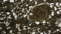
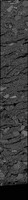
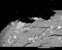
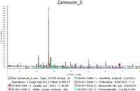
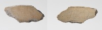
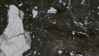
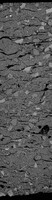
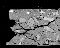
Backscattered Images acquired by Scanning Electron Microscopy at 50x and 200x magnification. Images from right to left top and bottom surface 1 to 4 at 50 x. Images from 5 to 13 at 200x.
Incisions description (parallel, curved and even impressions).
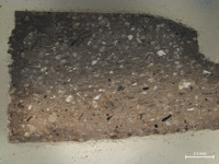
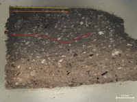
Inclusions characterization (color, shape and size).
Cross section characterization (in case of visible layers).
Backscattered Images acquired by Scanning Electron Microscopy at 50x and 200x magnification. Images from right to left top and bottom surface 1 to 7 at 50 x. Images from 8 to 20 at 200x.